Novrizal Saiful Basri, Margaretta Limawan, Odetta Natatilova, Rachmawati
Translator: Adrian Salim, Andrio Wishnu Prabowo, Arnetta Naomi L. Lalisang, Julistian, Muliyadi, Sony Sanjaya, Stefanny, Zamzania Anggia Shalih.
Surgery Department FMUI – Cipto Mangunkusumo Hospital, December 2011
CASE ILLUSTRATION
A 73 year old male came to the hospital with chief complaint of changes in urinary flow. He noticed weaker urinary flow with incomplete emptying of the bladder and occasional dribbling since two years prior hospital visit. There was history of red-colored urine, pain in the end of urination and an increased frequency of urination, especially at night. Assessment with IPSS showed a score of 27. There was no history of flank pain, cloudy urine, fever, nausea and vomiting. The patient had two episodes of stroke, but no history of diabetes mellitus and hypertension.
Physical examination demonstrated hypertension of 160/90 mmHg with no palpable mass or tenderness on Costo Vertebro Angle (CVA). No palpable mass and pain with palpation on the suprasymphisis area, bladder is empty. No stenosis was found at the external urethral orifice. Digital rectal examination showed normal resting anal sphincter tone, smooth and symmetrical prostate gland with firm consistency, no tenderness or nodule found. Prostate was estimated to weigh around 40 grams.
Laboratory tests showed normal levels of full blood count and kidney function, uric acid 9.4 mg/dl, total PSA 10.01 mg/dl. Urinalysis results are within normal values. Abdominal radiography revealed an apparent bladder stone of 35 x 25 mm , while sonography found bladder stone and left kidney stone. Uroflowmetry test showed Qmax value of 6.7 ml/sec; Q average 4.7 mL/sec; Void Volume 72 cc. Sonography of the prostate found the Rest Volume is 30 cc; Prostate Protrusion 9.59 mm; Prostate Size 110 cc (figure 1). Pathology report showed findings consistent with prostate hyperplasia.
The patient is then diagnosed with Benign Prostate Hyperplasia (BPH) and vesicolithiasis; and is planned to undergo cystoscopy and biopsy, sectio alta, and open prostatectomy.
Figure 1. Sonography of the Prostate
LITERATURE REVIEW
DEFINITION AND EPIDEMIOLOGY
Benign prostate hyperplasia (BPH) is a histopathologic term used to describe a true hyperplasia of the cells in the transitional zone of the prostate. The incidence of BPH is age-related with an estimated incidence of 70% in male above age 60 years; this number increases up to 90% in male above 80 years. Urology Department Cipto-Mangunkusumo Hospital (RSCM) reported 200-300 cases of BPH annually. While not immediately life-threatening, BPH may cause lower urinary tract symptoms (LUTS) that interfere with activity of daily living, thus significantly reduce patient’s quality of life.
DIAGNOSIS
Diagnosis of BPH is usually made using initial assessment with additional tests. One of the tool widely used to guide the identification of LUTS in BPH is the International Prostate Symptom Score (IPSS) (Figure 2). Digital rectal examination (DRE) may help assess the size and consistency of the prostate gland, while simultaneously detect any nodules that are suspicious for malignancy and assess anal sphincter tone and bulbocavernous reflex that indicate abnormalities in sacral reflex arc.

Figure 2. The Questionnaire for International Prostate Symptom Score (IPSS)
After the initial assessment, there are several tests available that help the diagnosis and management of BPH patients, including kidney function tests, prostate specific antigen (PSA) level, urinalysis, voiding diary, uroflowmetry, post voiding residual urine (PVR), urinary tract imaging, urethrocystoscopy, and urodynamic (pressure flow) study. The diagnosis of BPH usually consists of clinical examination (including DRE), urinalysis and sonography of the prostate.
MANAGEMENT
The goal of therapy in BPH focused mainly in improving patient’s quality of life. There are three different approaches available, each with its own modalities: (1) watchful waiting, (2) medical treaments, and (3) surgical intervention. Doctors will choose which approach is used based on the degree of symptoms, patient’s general health condition and the objective findings caused by the disease and other comorbidities.
The goal of therapy in BPH focused mainly in improving patient’s quality of life. There are three different approaches available, each with its own modalities: (1) watchful waiting, (2) medical treaments, and (3) surgical intervention. Doctors will choose which approach is used based on the degree of symptoms, patient’s general health condition and the objective findings caused by the disease and other comorbidities.
Watchful waiting is advised for patients with an IPSS score below 7, which indicates mild symptoms with no interference on activity of daily living. Patient will not get any medical or surgical intervention, but advised of what kind of changes that prompt immediate consult to doctor. Doctor will also monitor the patient closely for any changes in the severity of the signs and symptoms.
At some point, patients will need medication to help ease the symptoms. As a general rule, patients with IPSS score >7 will need medical treatment with/or other interventions. The goal of medical treatment is to (1) reduce the prostate smooth muscles resistance (dynamic component) and (2) reduce the size of the prostate (static component).
Surgical intervention can be classified into two groups: ablative technique of the prostate gland (open prostatectomy, TURP, TUIP, TUVP, laser prostatectomy) and instrumentation technique (interstitial laser coagulation, TUNA, TUMT, baloon dilatation, urethral stent).
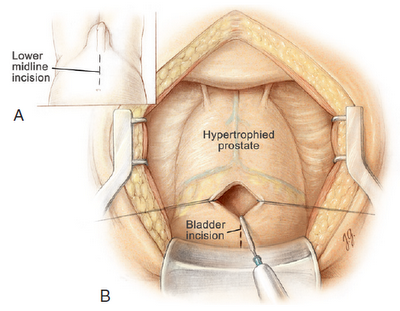
Figure 3. Open Prostatectomy with Suprapubic Approach
Open prostatectomy (figure 3) is the oldest, most invasive yet the most efficient way of surgical intervention with reported improvement of symptoms 98%. This open prostatectomy is done either with transvesical or retropubic approach. Prostate, internal urethral orifice and ureter orifice are then identified. Mucosa is then incised beside the internal urethral orifice at the 6 to 12 o’clock, followed by fracturation and prostate enucleation.
Glossary
Nocturia : [nox night + -uria] excessive urination at night
Nocturia : [nox night + -uria] excessive urination at night
Urgency : the sudden irresistable urge to void
Prostatectomy :[prostate + -ectomy] the removal of whole/part of the prostate gland
Cystoscopy : direct visual examination of the urinary tract using cystoscope
REFERENCES:
- Ikatan Ahli Urologi Indonesia. Pedoman Penatalaksanaan BPH di Indonesia. [Accessed at 20 Oktober 2011]. Available at: http://www.iaui.or.id/ast/file/bph.pdf.
- Roehrborn CG. Benign prostatic hyperplasia: etiology, pathophysiology, epidemiology, and natural history. In: Wein AJ, Kavoussi LR, Novick AC, Partin AW, Peters CA, editors. Campbell-Walsh Urology. 10th ed. Philadelphia: Elsevier Saunders; 2012. p. 2556-96
- Han M, Partin AW. Retropubic and suprapubic open prostatectomy. In: Wein AJ, Kavoussi LR, Novick AC, Partin AW, Peters CA, editors. Campbell-Walsh Urology. 10th ed. Philadelphia: Elsevier Saunders; 2012. p. 2695-703
- Syahputra FA, Umbas R. Diagnosis dan tatalaksana pembesaran prostat jinak: Peran antagonis reseptor adrenergik-α dan inhibitor 5-α reduktase. In: Birowo P, Syahputra FA, Ririmasse MP, Ismet MF, editor. Common Urologic Problems in Daily Primary Practice (CUPID) 2010. Ed 2. Jakarta: PLD FKUI dan Departemen Urologi FKUI RSCM; 2010. p.74-80.
- Rahardjo D. Prostat: kelainan-kelainan jinak, diagnosis dan penanganan. Jakarta: Sub Bagian Urologi Bagian Bedah FKUI; 1999. p.15-60.
- AUA practice guidelines committee. AUA guideline on management of benign prostatic hyperplasia. Chapter 1: diagnosis and treatment recommendations. American Urological Association 2010.
- Dorland, WA Newman. Kamus Kedokteran Dorland. Huriawati Hartanto et al, editor. 29thed. Jakarta: EGC; 2002.


